TriFoil Imaging – Preclinical Imaging Products
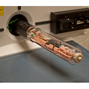
TriFoil Imaging is a leading developer and manufacturer of multi-modality nuclear and molecular imaging systems used in pre-clinical research. With the acquisition of Bioscan Inc.’s assets in 2013, TriFoil Imaging has added the development and manufacturing of tomographic optical imaging systems to their list of abilities.
With a wide variety of systems installations (over 300) TriFoil continues to drive product innovation in the in vivo molecular imaging market with its one-of-a-kind Triumph® tri-modality (PET-PET-CT) imaging system. TriFoil are soon to release the worlds first ever multi-modality benchtop series; the InSyTe imaging platforms. TriFoil Imaging’s technologies enable pharmaceutical companies to reduce costs and standardise analytical techniques across key areas of the drug development/discovery chain.
Products
 Their extensive range of technologies include:
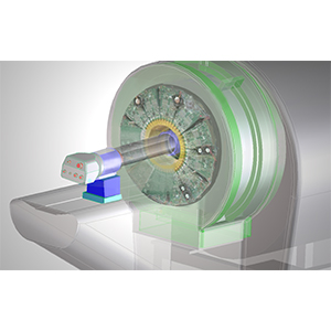
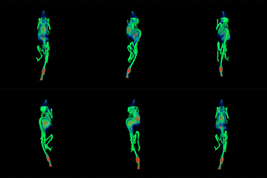
- Triumph® – The world’s first tri-modality system, offers the highest quality and best value by combining three modalities (PET, SPECT, and CT) all in one reliable and easy to use system.
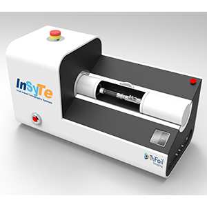
- InSyTeTM Series – First-ever table top imaging series to offer PET, SPECT or 3D Optical together with CT in a compact, easy to use package. These systems have the option to be purchased with a single modality at the initially and upgraded later to incorporate CT. It has a simple, consistent user- friendly interface for image reconstruction and co-registration across all modalities.
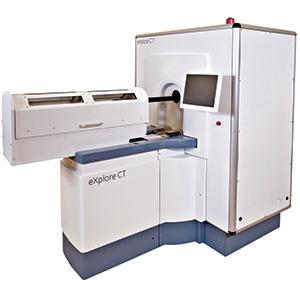
- eXplore CT 120 – A dedicated preclinical in vivo micro CT scanner designed for high-quality, high-throughput scanning in a wide variety of applications. It is the only CT system able to provide a true cardiac-gated imaging capability.
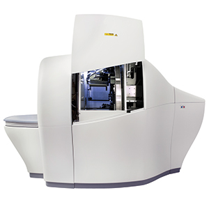
- Fluorescence Emission Computed Tomography (FLECT) – A revolutionary new optical imaging technology. It is the world’s first and only true tomographic Preclinical optical imaging system. The system is able to reconstructs staining pattern from the photons emission data from molecular markers acquired by sensitive detectors in a 360° arc around the animal. Then by utilising a state of the art 3D restorative algorithm based on an accurate model of light propagation in tissue, reconstruct the structure of the staining of the molecular markers. The data is equivalent to that generated by PET or SPECT systems.
