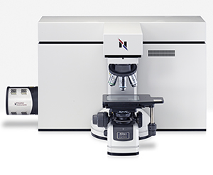
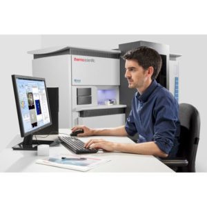
RISE Microscope – Raman Imaging and Scanning Electron Microscope
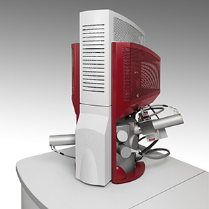
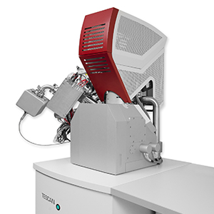
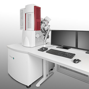
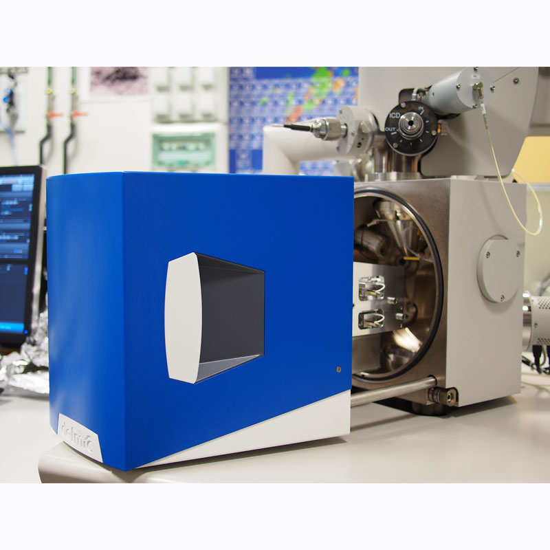
TESCAN and WITec leveraging on their respective expertise and experience have collaborated to create the worlds first fully-integrated Raman Imaging and Scanning Electron Microscope (RISE microscope). The RISE microscope incorporates and includes all the benefits of a stand-alone SEM and a top class confocal Raman imaging microscope, taking correlative microscopy to a new level.
RISE Microscope Features
The RISE microscope offers the following benefits:
- Quick and convenient switching been SEM and Raman modes
- Automated sample transfer from the Raman microscope to the SEM
- Integrated and easy-to-use software
- Correlation of the measurement results and image overlay
- No compromise from either the SEM or Raman system
- No risk of contamination going from SEM to Raman or vice versa
What Does it Offer?
RISE Microscopy offers correlative scanning electron and Raman imaging. It combines ultra-structural analysis via SEM, with molecular Raman imaging which provides chemical compound information resulting in comprehensive sample analysis.
Scanning Electron Microscope SEM
The SEM is a powerful tool for imaging samples at high magnification, while at the same time providing excellent resolution, all the way down to the nanoscale. It is able to yield information about the morphology, topography and chemical composition of the sample.
Combining the SEM with a focussed ion beam (FIB) source adds even more analytical capabilities. TESCAN are able to supply thermal emission SEMs and FEG-SEMs with FIBs. All TESCAN systems benefit from modern optics with unique Wide Field Optics design and their proprietary intermediate lens (IML) in addition to a conventional lens which allow numerous working and displaying modes. Other features common to TESCAN SEMs include:
- Ultra-fast scanning
- In-Flight Beam Tracing
- Automated procedures and user-friendly software
- In-Beam secondary and backscattered electron detectors
Confocal Raman Imaging
WITecs Confocal Raman Microscopy and Imaging System combines Raman spectroscopy and confocal microscopy. This enables Raman imaging with the information of a complete Raman spectrum at every image pixel and lateral resolution at the limit of diffraction (~200nm).
WITecs expertise in this area allows them to offer:
- Raman imaging with unprecedented performance in speed, sensitivity and resolution
- Outstanding depth resolution, perfect for 3D imaging and depth profiling
- Excellent sensitivity and performance in spectral resolution thanks to the high-throughput lens-based stereoscopic system
- Ultra-fast Raman imaging option, with 0.76ms integration time per spectrum possible
- Non-destructive imaging and no sample preparation necessary
Raman Imaging
Raman imaging can provide the following information:
- Peak Intensity Quantity/amount of a specific compound
- Peak Shift Identification of stress and strain states
- Peak Width Degree of crystallinity
- Polarisation State Crystal symmetry and orientation
Applications
RISE microscopy is suited to applications such as:
- Materials science
- Nanotechnology
- Life Science
- Pharmaceuticals
- Polymer science
- Geoscience
Graphene

Left – Graphene sample imaged using SEM
Middle – Colour-coded confocal Raman image. The colours show the graphene layers and wrinkles. The image is 20µm x 20µm, 150 x 150 pixels = 22500 spectra. Integration time of 0.05 s/spectra.
Right – SEM image overlaid with confocal Raman image.

 Download the AXT Life Science Product Guide