Lyncean Technologies – Compact Light Sources – The Synchrotron Alternative
Lyncean Technologies, based in Fremont California was founded in 2002 to develop and commercialise the Compact Light Source (CLS), a miniature alternative to synchrotron facilities. Their technology originated from research carried out at the SLAC National Accelerator Laboratory (one of 10 Department of Energy (DOE) Office of Science laboratories) and Stanford University.
The Compact Light Source
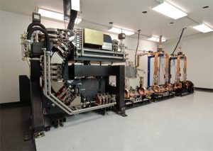
The Compact Light Source is a breakthrough technology that enables access to synchrotron-like analytical facilities in a homelab-type scenario. With researchers continuing to push the boundaries of science and probing materials and structures at the nanoscale, there is an increasing requirement for analytical X-rays such as those available at synchrotrons. In fact, the existing synchrotron in Australia is oversubscribed and having another similar facility, in a city other than Melbourne would be a great benefit to Australian researchers and industry.
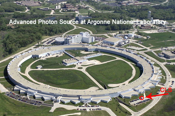 Synchrotrons are typically the size of football fields. As such they require dedicated facilities. The Lyncean CLS is some 200 times smaller and can be located in a typical laboratory room. As a result of the more compact design, the CLS does not require a team of dedicated engineers and technicians to run it and has been designed to be operated by academics and industrial end-users. Furthermore, it allows researchers to have a more accessible local facility
Lyncean have been able to achieve this massive reduction in size by replacing the conventional undulator magnets in the synchrotron design with laser undulator technology. The CLS produces a narrow beam of almost monochromatic X-rays that are adjustable in energy.
The Lyncean CLS generates a tuneable, monochromatic, spatially coherent beam that can be either:
- Focussed for general diffraction experiments e.g. powder diffraction or SAXS
- Unfocussed for imaging of large samples
The large area beam is able to provide high resolution tomographic imaging of biological specimens. The nature of the beam enables new methods of imaging development beyond conventional radiography opening the doors for new possibilities in medical research.
The first CLS has been installed at the Center for Advanced Laser Applications in Germany, a joint project of the Ludwig Maximilians University of Munich and the Technical University Munich (TUM). This system has been in routine operation since April 2015.
Applications
The CLS is suited to a host of conventional X-ray applications as well as many new ones that are outside the capabilities of homelab X-ray systems. Some example applications include:
- Protein crystallography
- Drug development
- Phase contrast imaging
- Micro tomography
- Powder diffraction
- Pharmaceuticals
- Structural biology
- Small angle scattering
- Nanotechnology
- Advanced materials research
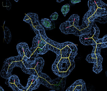
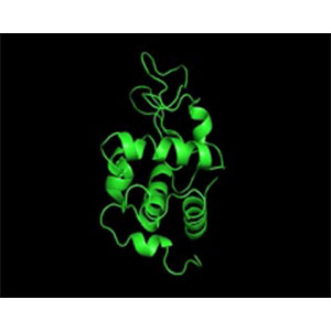
Electron density map from refined lysozyme model. Unmodeled water molecules are visible in the upper portion of the picture.
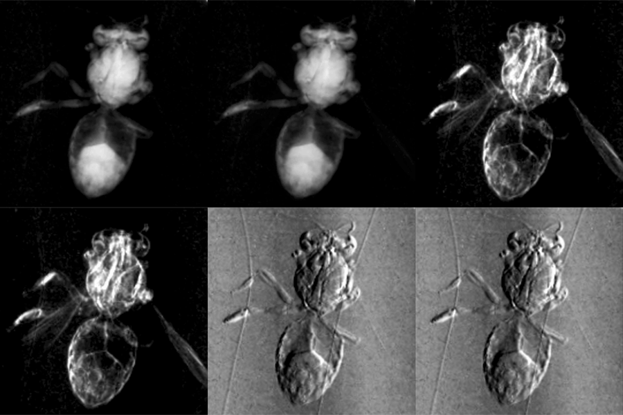
CLS phase contrast images of a bee and a moth. First image is absorption, second is dark field (local scattering intensity), and third is the phase image. (Images courtesy of Bech, M., Bunk, O., David, C., Ruth, R., Rifkin, J., Loewen, R., Feidenhans’l, R. & Pfeiffer, F. (2009). J. Synchrotron Rad. 16, 43-47)
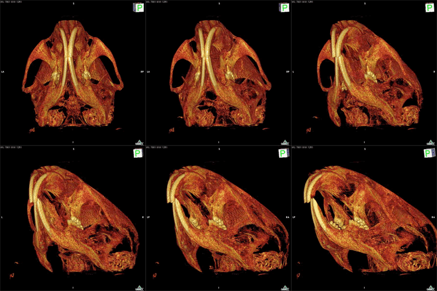
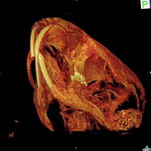
A high resolution 3D rendering of a mouse head created using a newly developed Rayonix CCD taken on the CLS (2010). (Video courtesy of BioMedical Physics group, Prof. F. Pfeiffer, TU München)

