Fluorescence and Holographic Tomographic Microscopy for Live Cell Imaging – 3D Cell Explorer-Fluo
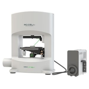
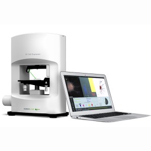
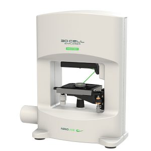
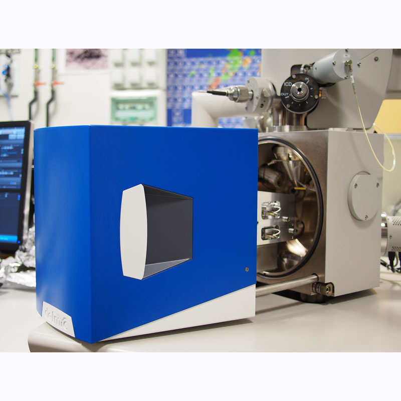
The Nanolive 3D Cell Explorer-fluo is the most powerful imaging tool for investigating the behaviour of live cells providing correlative microscopy capabilities. It adds functional information gained by fluorescence microscopy to the non-invasive 3D holographic tomographic capabilities of the 3D Cell Explorer generating data to provide a more comprehensive understanding of how cells and subcellular components really respond to treatments and therapies.
Multi-channel fluorescence microscopy is a well-respected technique in biology and its incorporation into the 3DCX platform opens up new possibilities in scientific research and live cell imaging.
Fluorescence Microscopy
Fluorescence microscopy allows biological and biomedical science researchers to visualise specific cell compartments or features by staining them using various fluorescent probes. In this way, details can be revealed that would not otherwise be visible using other traditional modes. It is popular due to its high sensitivity and specificity.
The fluorescent probes allow you to identify specific subcellular components. Using multiple fluorescent labels allows you to simultaneously identify several components at the same time amongst a matrix of non-fluorescent material. This enables you to get a better understanding of the internal structure of your cells and how they operate.
Holographic Tomographic Imaging
Using the physical characteristic of refractive index to differentiate subcellular components, the 3DCX is able to produce detailed 3D images of cells in real time revealing physical and structural features. In doing so, long terms 4D studies can be carried out with images captured every second to produce dynamic behavioural studies of cells.
The Best of Both Worlds
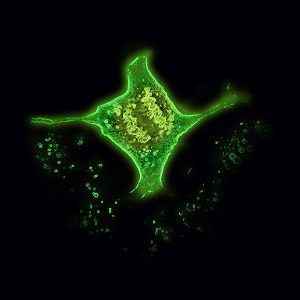
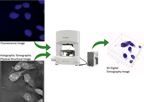
The 3D Cell Explorer- fluo allows you to generate holographic tomographic and fluorescence data sets and combine these together, thus merging chemical imaging (from the fluorescence microscope) with physical and structural information (from the holographic tomographic microscope). By overlaying these two datasets, you can transform 2D fluorescence data into 3D cell tomography to produce correlative results which allows you to see how various individual subcellular units are behaving. Using up to 10 different fluorescent markers in parallel you can effectively look at 10 different cellular components at the same time.
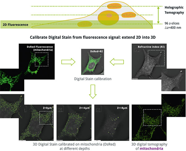
By combining fluorescence and holographic tomography, you can localise organelles at different cell depths e.g. mitochondria. This enables you to image your cells in a whole new dimension, introducing an enhanced level of understanding.
Explore 10 Markers in Parallel
The Nanolive 3DCX-fluo is the first microscope that allows the user to image as many as 10 channels simultaneously. This really enables you to accelerate your research, especially when used in conjunction with tomographic holography.
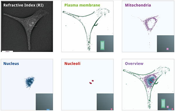
Digital stains calibrated for various organelles in mouse melanoma skin cancer cell (B16).
Reduce Artifacts and Errors
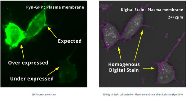
By combining the power of fluorescence and holographic tomographic microscopy you can reduce imaging artifacts and produce more reliable cellular measurements, thus generating more accurate data.
Specifications
| Holographic Tomography | Fluorescence Imaging | |
|---|---|---|
| Illumination Source | Class 1 laser low power (λ=520 nm, sample exposure 0.2 mW/mm2) | High speed switchable <100 μs Lifetime >20,000 hours each channel |
| Resolution (nm) | x,y: 200; z: 400 | x,y: 400 |
| Field-of-View (µm) | 90 x 90 x 30 | 100 x 100 |
| Channels | Up to 9 simultaneous | 3: DAPI (blue), GFP (green), TRITC (red) |
| Imaging | 3D / 4D (time-lapse) | 2D |
| Time Resolution | 0.6 fps 3D RI frame | 5 fps each channel |
| Camera | CMOS sensor – 73 dB of dynamic range / Quantum efficiency of 70 % Readout noise: 6.6 e– | |
| Dimensions (cm) | 38 x 45 x 17 | 21 x 23 x 10 |
| Weight (kg) | 8 | 4 |

