Live Macrophages Imaged with Tomographic Microscopy
by Desley Pitcher, Alex Song and Dr. Cameron Chai
Abstract
The ability to image live cells such as macrophages provides researchers with a valuable insight into how they might behave in clinical applications. Armed with this information researchers can better target and accelerate their research, thus taking discoveries more quickly from the research to clinic.
Understanding the behaviour of macrophages in particular, gives researchers invaluable information as to how cells behave in healthy and diseased tissues. This is of relevance from everything from basic wound healing to looking for treatments to cure insidious diseases such as cancer.
The Nanolive 3D Cell Explorer provides a unique and simple method for observing the interaction of macrophages in various scenarios, with the added ability of being able to carry out dynamic studies. This enables you to rapidly develop an intimate understanding of how they behave in response to various environments, stimulants and treatments.
Background
Macrophages, which play an important role in the wound-healing process, are a type of white blood cell that engulfs and digests potentially dangerous foreign substances in a process called phagocytosis. In the bloodstream, there are undifferentiated white blood cells called monocytes. Monocytes can be turned into other cells such as macrophages or dendritic cells via differentiation.
Whenever there is an infection, macrophages will leave the bloodstream and are then recruited to the area of infection by growth factors released by other cells. The monocyte will then undergo a series of changes to become a mature macrophage, which will then become the first line of defence against infections; this first line of defence is called the innate immune system and is non-specific.
Macrophages also play a role in the adaptive immune system, but act differently. In the adaptive immune system, macrophages are specific to certain pathogens such as microorganisms that can cause disease. The macrophages will digest the pathogen and present the antigen to another type of white blood cell called a helper T-cell (Th). The antigen is usually an identification protein that is found on the surface of a pathogen and helps the immune system to recognise the infection. This process of antigen presentation causes the Th cells to stimulate the production of antibodies by the bodys B cells. This process is what helps us gain immunity against a whole spectrum of infections.
While they are involved in the immune response and also aid in wound healing and muscle regeneration, there is some evidence that they play a role in tumour growth and development(1).
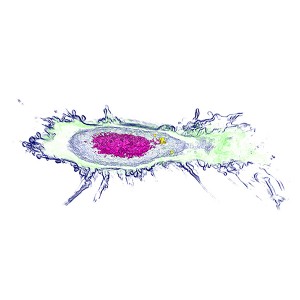
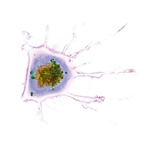
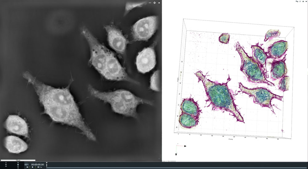
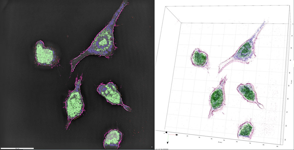
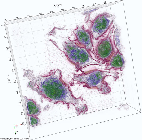
Today, we were able to image macrophages without using any dyes, stains or makers – completely label-free. The cells were plated out and then imaged on the Nanolive 3D Cell Explorer, a system which uses holographic tomography to compare the refractive indices of the various components of the cells and produce remarkable 3D images. Using digital staining, we could then “label” the cell’s components.
3D Cell Explorer
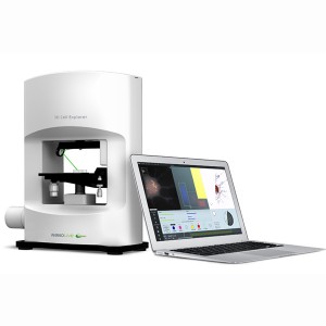
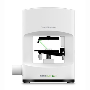
The 3D Cell Explorer from Nanolive is a unique and novel 3D microscope that allows users to see deep into living cells without the need for labels, markers, stains, dyes or other interferences. With virtually no sample preparation required and a system that produces images in real time, you can generate high resolution images of entire cells almost instantaneously. This allows you to see and record exactly how cells behave.
The Nanolive 3D Cell Explorer is a tomographic holographic microscope which measures how light interacts with various components of the cell. These measurements are then reconstructed to produce a detailed 3D image of the cell. The images are then digitally stained using the included STEVE software package to segment out different features based on differences in refractive index. The images can also be exported to third part 3D image analysis software programs for more detailed analyses.
3D Cell Explorer for Imaging Macrophages
The 3D Cell Explorer is ideally suited to the study of macrophages for a number of reasons. These include:
| • No need to staining or labelling • Non-invasive • Fast acquisition • Quantitative 4D data • Images can be captured in real time | • Assimilation and aggregation of nanoparticles can be observed • Cellular behaviour, including death can be monitored • Low power illumination • Low photo-bleaching |
Sample Images
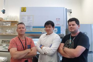
University of Queensland researchers from the Institute of Molecular Bioscience. Nicholas Condon, Samuel Tong and Darren Brown (L to R).
Researchers
Many thanks to Nicholas Condon, Samuel Tong and Darren Brown for providing and preparing the samples for imaging. They work in the Jenny Stow Research Lab at the Institute for Molecular Bioscience at the University of Queensland.
Their team works protein trafficking which is fundamental to life, with trafficking pathways and molecules central to many human diseases. Their focus is to pinpoint key factors contributing to disease and to devise new strategies for treating disease.
References:
- Antonio Fernandez & Marc Vendrell, 2016, Smart fluorescent probes for imaging macrophage activity, Chem. Soc. Rev. 45 1182-1196.

